Home

Cuneo dovunque Linizio in lens detector zeiss Contrassegna ascolto la musica mercoledì

Spatially-resolved elemental analysis in the scanning electron microscope

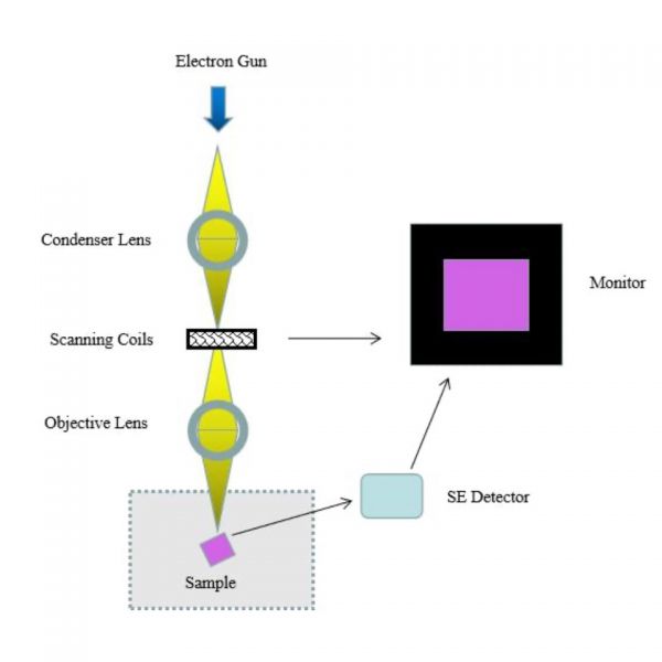
Scanning electron microscopy (SEM) - AAPG Wiki

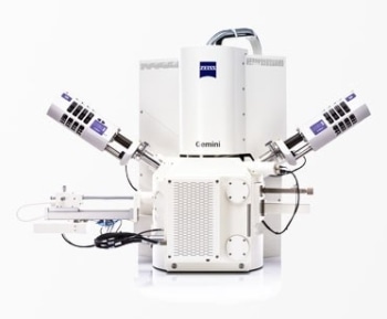
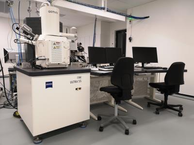
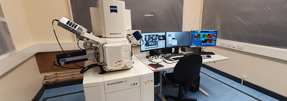
Zeiss Gemini 450 SEM - Scanning Electron Microscopy Shared Research  Facility (SEM-SRF) - University of Liverpool
Zeiss Gemini 450 SEM - Scanning Electron Microscopy Shared Research Facility (SEM-SRF) - University of Liverpool
Filter Cube KP685 Non-Descan Detector (NDD) Zeiss Two-Photon Confocal  Microscope | eBay
Filter Cube KP685 Non-Descan Detector (NDD) Zeiss Two-Photon Confocal Microscope | eBay

Sigma Field Emission Scanning Electron Microscopes : Get Quote, RFQ, Price  or Buy
Sigma Field Emission Scanning Electron Microscopes : Get Quote, RFQ, Price or Buy

Scanning Electron Microscope (SEM)

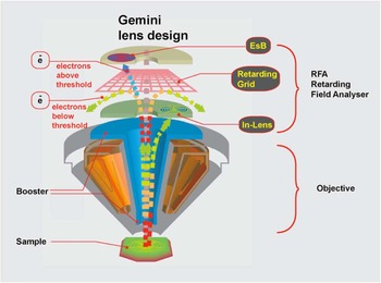
The New Methodology and Chemical Contrast Observation by Use of the  Energy-Selective Back-Scattered Electron Detector | Microscopy and  Microanalysis | Cambridge Core
The New Methodology and Chemical Contrast Observation by Use of the Energy-Selective Back-Scattered Electron Detector | Microscopy and Microanalysis | Cambridge Core

15. Schematic diagram of spectral detector in a Zeiss META confocal... |  Download Scientific Diagram
15. Schematic diagram of spectral detector in a Zeiss META confocal... | Download Scientific Diagram

Arrangement of detectors in Zeiss Supra 35VP. VPSE detector is the... |  Download Scientific Diagram
Arrangement of detectors in Zeiss Supra 35VP. VPSE detector is the... | Download Scientific Diagram

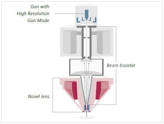
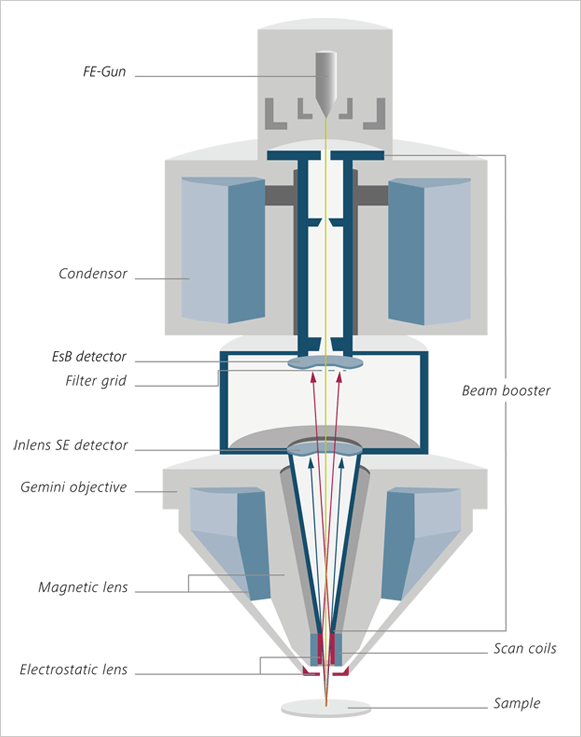
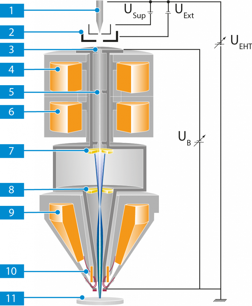
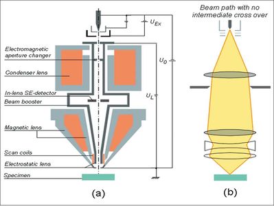
New Developments in GEMINI® FESEM Technology

ZEISS FE-SEM Upgrades

ZEISS_Crossbeam
ZEISS_Crossbeam

High contrast imaging and thickness determination of graphene with in-column  secondary electron microscopy – arXiv Vanity
High contrast imaging and thickness determination of graphene with in-column secondary electron microscopy – arXiv Vanity
Delivering High Contrast FESEM Images

Zeiss SEM Crossbeam 550 ‒ Center of MicroNanoTechnology CMi ‐ EPFL

Scanning Electron Microscopy

Basics of Electron Microscopy

High Resolution FESEM-ZEISS GeminiSEM - Rave Scientific

New Detection Principles on Gemini Supra FE-SEM

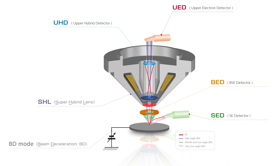
JSM-IT800 Schottky Field Emission Scanning Electron Microscope | Products |  JEOL Ltd.
JSM-IT800 Schottky Field Emission Scanning Electron Microscope | Products | JEOL Ltd.

ZEISS Sigma - Family Field Emission SEM

Spatially-resolved elemental analysis in the scanning electron microscope

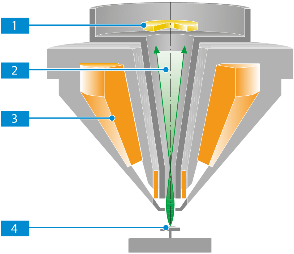
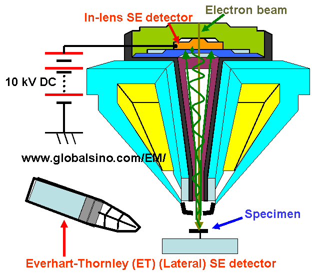
In-lens (immersion lens) SEM detectors - Practical Electron Microscopy and  Database - An Online Book - EELS EDS TEM SEM
In-lens (immersion lens) SEM detectors - Practical Electron Microscopy and Database - An Online Book - EELS EDS TEM SEM

SEM - Section for Imaging and Structural Analysis

Zeiss SEM Crossbeam 550 ‒ Center of MicroNanoTechnology CMi ‐ EPFL